
Objective
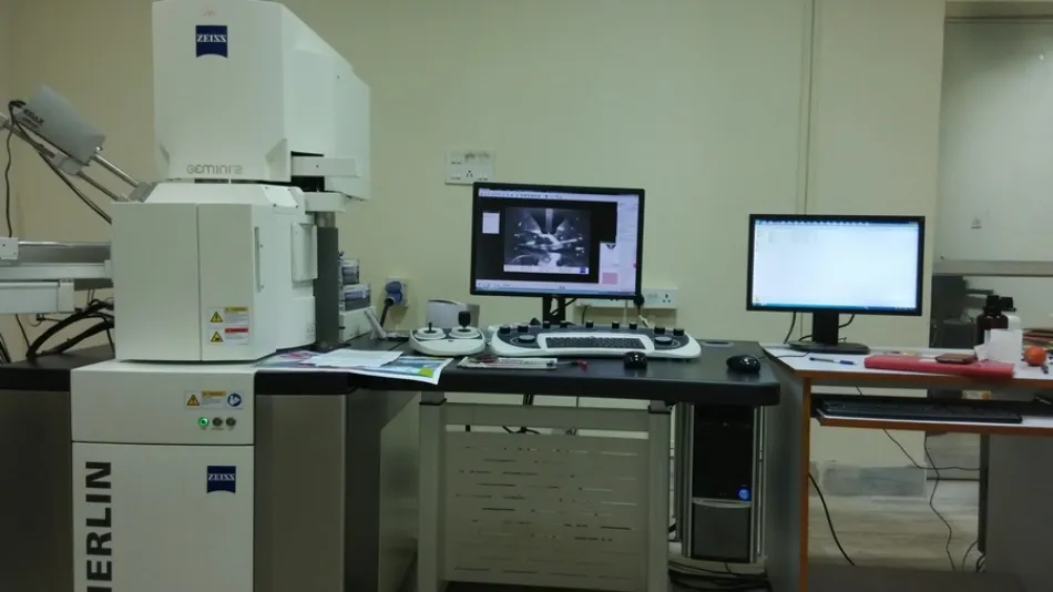
Merlin is a field emission gun scanning electron microscope. This machine is used to see the morphology of different samples. MERLIN with the GEMINI II column combines ultra fast analytics, high resolution imaging using advanced detection modes, and future assured configuration flexibility on one single system.
Sample Details
1. In order to observed with an electron microscope sample should be conductive (for non conductive samples you have to do very thin conductive coating upon the sample).
2. Sample must be free from any type of solvent (water, oil etc) free otherwise it will damage the system vacuum.
3. For powder samples very small amount is needed, for thin films 1cm×1cm is enough, for solid samples 2cm×2cm×2cm is more than sufficient.
2. Sample must be free from any type of solvent (water, oil etc) free otherwise it will damage the system vacuum.
3. For powder samples very small amount is needed, for thin films 1cm×1cm is enough, for solid samples 2cm×2cm×2cm is more than sufficient.
Utility and Working Principle
FESEM is the abbreviation of Field Emission Scanning Electron Microscope. FESEM is a microscope that works with electron instead of light. These electrons are liberated from a field emission source and accelerate in a high electric field gradient. Within the high vacuum column these so called primary electron are focused and deflected by electronic lenses to produce a narrow scan beam that bombards upon the object (sample). As a result secondary electrons are emitted from each spot of the object. The angle and the velocity of the electrons (secondary) relates to the surface structure of the object. A detector catches these secondary electrons and produces an electronic signal. This signal is amplified and transformed to a video scan image that can be seen on monitor.